Pleuropneumonia is the term applied to pneumonia when it extends from the main airways (bronchopneumonia) to the thin membrane on the surface of the lung (the pleura) and the surrounding thoracic cavity. Once a bacterial pneumonia reaches the pleura, horses respond by producing large volumes of proteinaceous fluid. Pleuropneumonia is the most severe form of pneumonia and often requires long-term hospitalization and intensive care to resolve. Horses with pleuropneumonia often have endotoxemia, thus they can develop many complications, including laminitis and death.
Pleuropneumonia is most commonly associated with long-distance transportation (shipping fever), but it also develops following esophageal obstruction (choke) and secondary aspiration pneumonia. Stress is often considered a contributing cause of shipping fever because travel is known to weaken host immune responses against infection. However, the position of the horse’s head and the way they are fed during travel likely play a more important role.
Normally, horses have their heads down for long periods of time, which allow them to easily clear debris from their trachea and lower airways. Horses that are transported with their heads tied in elevated positions have decreased ability to clear their airways. In addition to head position, hay is often placed in a bag directly in front of the horse’s nose, so they are constantly inhaling debris and bacteria that then settles in the lower airways and incites inflammation and infection.
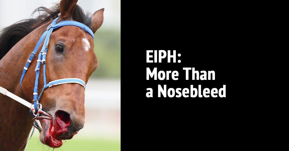
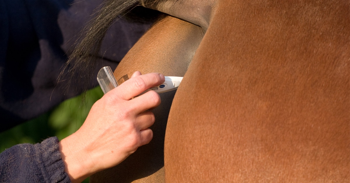
Pleuropneumonia does not always develop immediately and can go unnoticed for days to weeks after the inciting event. Obvious clinical signs include cough, increased respiratory rate and effort and purulent (pus-like) nasal discharge coming from both nostrils that often has a fetid odor. Subtle signs include fever (greater than 101.5°F), weight loss, decreased appetite and malaise. It is important to note that horses with pleuropneumonia do not always develop nasal discharge or cough. Diagnosis of pleuropneumonia is confirmed via imaging of the lungs and sampling of the airways to identify bacterial infection. Ultrasound is the most frequently employed mode of imaging, as it is portable and can be easily performed in ambulatory practice. Ultrasound can identify and measure fluid in the thoracic cavity, which is the hallmark of pleuropneumonia. Ultrasonography can only be used to image the surface of the lung, so radiography may also be performed to assess the deeper lung tissue.
Once pleuropneumonia has been identified via ultrasound, pleural fluid can be sampled for bacterial culture and microscopic evaluation. Bacterial culture and antimicrobial susceptibility testing ensure that horses will be treated with targeted antimicrobial therapy. In normal horses, the mediastinum (the division between the right and left side of the thoracic cavity) has small slits in it that allow movement of small amounts of fluid from one side of the thorax to the other. In pleuropneumonia, large amounts of inflammatory materials (fibrin, leukocytes and cellular debris) obstruct these openings so there is complete separation of the two sides. Once this has occurred, it is possible for different bacteria to cause infections in the separate thoracic spaces, thus samples should be taken from both sides. In addition to sampling pleural fluid, a transtracheal wash should also be performed and the fluid cultured for bacteria. Adjunct diagnostics may be utilized to assess a horse’s systemic health and include a complete blood count, measurement of acute phase proteins (e.g., serum amyloid A), chemistry panel and blood gas analysis.
The pillars of therapy for pleuropneumonia include drainage of pleural fluid, antimicrobial therapy and supportive care. Indwelling chest tubes may be placed to facilitate constant drainage and allow for lavage and administration of intrathoracic medication. Intravenous antimicrobial administration is preferred over oral or intramuscular routes to reach higher drug levels in diseased tissue.
Antimicrobials may also be nebulized to increase penetration into the lower airways while avoiding potential adverse systemic effects. Large volumes of fluid can be lost each day from the thoracic cavity, therefore intravenous fluid therapy is often required to ensure adequate hydration. Non-steroidal anti-inflammatory agents are administered to reduce inflammation, attenuate endotoxemia and provide analgesia. Supplemental oxygen, bronchodilators, intravenous nutrition, plasma transfusions, cryotherapy for laminitis prevention and surgical intervention may also be warranted. As the infection begins to resolve and lungs heal, horses can be transitioned from injectable to oral antimicrobials.
Many horses are discharged for long-term antimicrobial therapy at home, and treatment may be necessary for multiple months. Once pleuropneumonia has resolved, horses can slowly return to work, and many go back to their previous level of performance.
The Latest